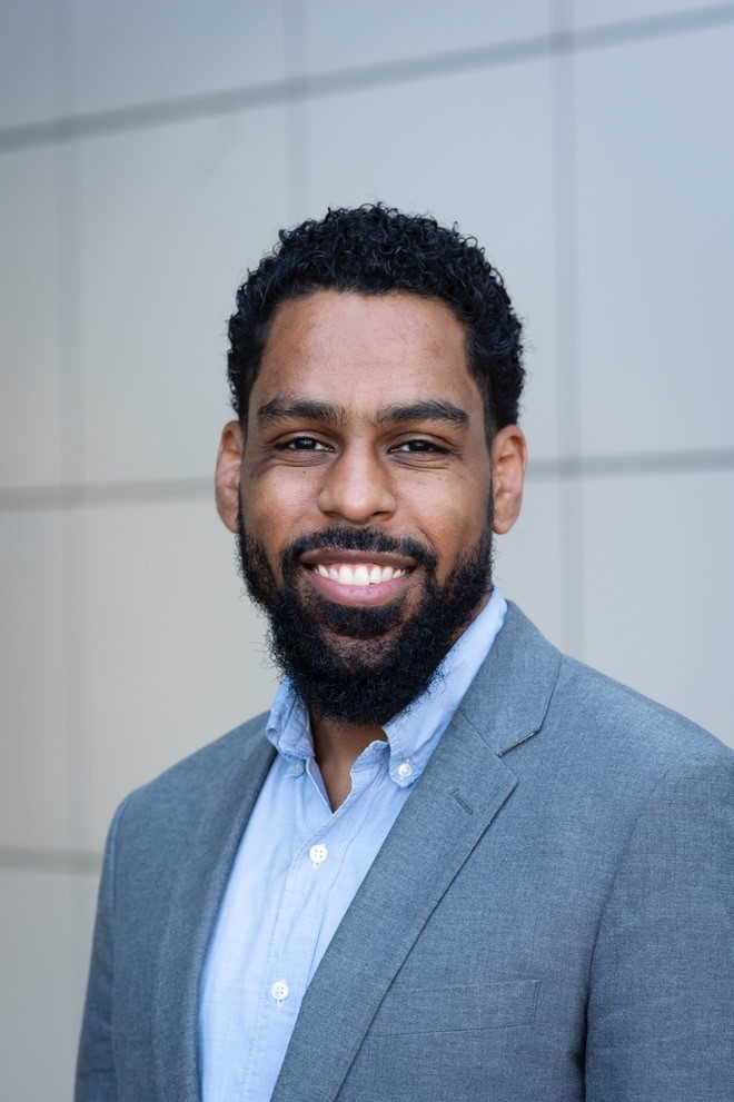
Karl Lewis, Cornell University – Novel Approaches to Study Early Cell Changes in Musculoskeletal Disease
 On Cornell University College of Engineering Week: How do we better understand cell communication?
Karl Lewis, assistant professor of biomedical engineering, deciphers the signals.
Dr. Karl Lewis is an assistant professor at the Meinig School of Biomedical Engineering at Cornell University. Dr. Lewis’s research interests center on understanding the interplay of mechanical cues and biological changes in musculoskeletal tissues. His group develops novel intravital imaging techniques for studying mechanotransduction and mechanobiology in vivo. They interrogate how acute sensing mechanisms in musculoskeletal cells relate to tissue-level changes in healthy and disease states, with the objective to use this knowledge to identify new targets for therapeutic intervention in musculoskeletal disease. Moreover, the Lewis Lab is dedicated to expanding analysis techniques for histological and live imaging data.
Novel Approaches to Study Early Cell Changes in Musculoskeletal Disease
Just like humans wink and smile, cells have different forms of communication. One particular type of communication, calcium ion signaling, could help scientists unlock secrets about tissue diseases such as arthritis and osteoporosis – that is, if we can understand the signals.
Calcium ion signals are very fast fluctuations in calcium content in cells that communicate biological events, such as a response to your body being loaded with weight. If you pick up a heavy box, the stress on your bone, cartilage, and tendons triggers the signaling, which in turn tells other cells how to respond. And unlike most molecular targets, calcium ion signaling is special because scientists can monitor it in real time and in living cells using basic microscopic approaches.
Given that we can apply load in a reliable and highly precise manner to bone, cartilage, and tendons in laboratory settings, these types of tissue make ideal model systems for studying calcium ion signaling. The challenge is that these signals are very fast, occurring in just milliseconds, and they occur at very small length scales in dense tissues.
My lab specializes in using an advanced type of microscopy to observe tissue cells in living organisms. We combine this imaging technique with custom designed loading devices to study signaling dynamics in real time. These types of studies offer the ability to see those very fast signals as changes in the cell’s conditions occur. We are a part of a burgeoning field where scientists are connecting what a cell is experiencing in its natural environment to what a cell is signaling.
The relationship between very short-term cellular activity and long-term tissue disease outcomes is not presently well established for many tissues, but if we can continue to learn from calcium ion signaling and what cells are trying communicate, we improve personalized and preventative medicine dramatically. Just like any good relationship, communication is key.


