 On Cornell University College of Engineering Week: The machinery in our bodies that creates bones or teeth can sometimes go awry.
Lara Estroff, professor and chair of the department of materials science and engineering, determines what we can do to combat this.
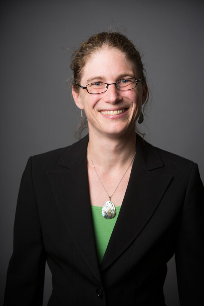
Lara Estroff is a professor and chair of the Department of Materials Science and Engineering at Cornell University. Her research focuses on bio-inspired materials synthesis, in particular, the study of crystal growth mechanisms in gels and their relationships to biomineralization. Among other honors, she is the recipient of the NSF Early Faculty Career Award and the J.D. Watson Young Investigator’s Award.
Pathological Mineralization
Anyone who has marveled at the intricacies of the lace-like details in a sand dollar shell or the smooth, curved surfaces of a deer antler, has experienced the beauty of biomineralization, the processes by which biological organisms create hard tissues.
In our own bodies, our teeth and bones are engineering masterpieces of structural elements grown by our cells to perform specific functions such as grinding our food and supporting our weight. Sometimes, however, the machinery responsible for mineralization misfunctions, and mineral ends up forming in tissues where it shouldn’t be. When this happens, we call it pathological mineralization.
Some better known examples of pathological mineralization are kidney stones and calcific aortic valve disease. In these diseases, research is focused on understanding the origins of the misfunctioning “machinery” as well as preventing the unwanted minerals from forming.
In my own lab, we study microcalcifications, small calcium-rich deposits, associated with breast tumors. We are interested in applying what we know from the study of healthy biomineralization to better understand the origins of microcalcifications in cancers. Specifically, we image tissue biopsies to create maps of the composition and morphology of individual calcifications and their surroundings. Using this information, we ask the questions: What was the local chemistry in the tissue within which the crystals grew? From this information, what can we learn about the disease progression and severity? And in the future, how can we use this information to inform treatment options?
We are particularly excited about this approach because the advanced imaging technique we use could be integrated into a modern pathology lab and used alongside current gold-standards for informing medical decisions, such as, when to treat the cancer with surgery versus other therapies.
